Ultrasound Case: Acute Cholangitis
A 72-year-old male presents with high-grade fever, severe jaundice, and right upper quadrant (RUQ) abdominal pain.
Case Presentation
History
- Chief Complaint: Fever, jaundice, and right upper quadrant (RUQ) pain (Charcot’s triad) progressing over 48 hours.
- History of Present Illness: * Colicky RUQ pain radiating to back.
- High-grade fever (39.2°C) with chills.
- Progressive yellowish discoloration of skin.
- Dark urine and pale stools.
- Past Medical History: Known gallstones, Type 2 diabetes mellitus, hypertension.
- Medications: Metformin, lisinopril.
- Laboratory Findings:
- WBC 19.5 x10³/µL with 90% neutrophils.
- Total bilirubin 5.2 mg/dL (direct 4.3).
- ALP 520 U/L, GGT 380 U/L.
- AST 180 U/L, ALT 160 U/L.
Physical Examination
- Vital Signs: Temp 39.1°C, HR 112, BP 90/60, RR 22.
- General: Icteric sclerae, dry mucous membranes.
- Abdomen: Tenderness in RUQ with guarding, positive Murphy’s sign. No rebound tenderness, no palpable masses.
- Neurologic: Mild confusion (new onset).
Clinical Suspicion: Given the history of gallstones and characteristic clinical presentation matching Charcot’s triad, acute ascending cholangitis with sepsis is highly suspected. Urgent ultrasound is requested to evaluate for biliary tree obstruction, confirm ductal dilation, and guide emergent decompression management.
Ultrasound Findings
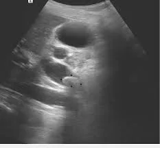
- Bile duct dilation: Common bile duct diameter measures 12mm; intrahepatic ducts show a >2mm diameter, yielding the “too many tubes” sign at the porta hepatis.
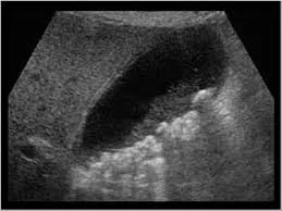
- Obstructing lesion: A 9mm hyperechoic stone is visualized in the distal CBD displaying clear posterior acoustic shadowing alongside proximal ductal dilation.
- Gallbladder findings: Multiple small gallstones are seen within the lumen; the gallbladder wall measures 4mm in thickness, showing mild reactive edema.
1. CBD dilation and choledocholithiasis: Common bile duct dilated to 12mm with an obstructing 9mm calculus demonstrating distinct posterior acoustic shadowing.
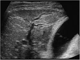
2. Intrahepatic duct dilation: Markedly dilated branching intrahepatic biliary ducts producing a classic 'too many tubes' sign.
3. Multiple gallstones: Acoustic-shadowing intraluminal gallbladder stones accompanied by reactive wall thickening.
Final Diagnosis: Acute Obstructive Cholangitis Secondary to Choledocholithiasis > Confirmed based on matching clinical findings (fever, jaundice, RUQ pain, confusion, hypotension) alongside key ultrasound features (extrahepatic obstructing stone, severe CBD dilation, and upstream intrahepatic duct ectasia).
Differential Diagnosis
- Acute Cholecystitis: Presents with GB wall thickening (>3mm) and pericholecystic fluid, but characteristically lacks central bile duct dilation or an extrahepatic obstructing calculus.
- Viral Hepatitis: Features diffuse liver heterogeneity and acute clinical jaundice, but lacks any mechanical ductal dilation or mobile intraluminal shadowing stones.
- Pancreatitis: Displays prominent pancreatic edema and peripancreatic fluid; however, it can coexist with secondary CBD dilation if a stone passes through the ampulla.
- Biliary Stricture: Features focal duct narrowing and gradual symptom onset, but typically presents without acute systemic infectious signs like high fevers and shaking chills.
- Cholangiocarcinoma: Presents with a solid mass lesion at the duct level or abrupt, malignant duct cutoff without clean, mobile acoustic-shadowing stones.