Liver Ultrasound
Liver ultrasound remains one of the most commonly performed abdominal imaging studies, offering a non-invasive, radiation-free method to evaluate hepatic anatomy and pathology. This comprehensive guide provides a systematic approach to liver ultrasound, covering essential technical aspects, normal anatomical features, and common pathological findings.
Liver anatomy
The liver is located in the right upper quadrant. It is almost completely covered by visceral peritoneum and is completely covered by a dense irregular connective tissue layer that lies deep to the peritoneum.
The liver is divided into two principal lobes (a large right lobe and a smaller left lobe) by the falciform ligament, a fold of the mesentery.
The falciform ligament extends from the undersurface of the diaphragm between the two principal lobes of the liver to the superior surface of the liver, helping to suspend the liver in the abdominal cavity.
In the free border of the falciform ligament is the ligamentum teres (round ligament), a remnant of the umbilical vein of the fetus; this fibrous cord extends from the liver to the umbilicus. The right and left coronary ligaments are narrow extensions of the parietal peritoneum that suspend the liver from the diaphragm.
Clinical Indications
Liver ultrasound is typically requested for the following clinical scenarios:
- Abnormal Liver Function Tests
- Evaluation of elevated ALT, AST, ALP, or bilirubin to assess for hepatitis, obstruction, or infiltrative diseases.
- Right Upper Quadrant Pain.
- Investigation of biliary colic, cholecystitis, hepatitis, or other hepatic pathologies.
- Hepatomegaly
- Assessment of suspected liver enlargement detected on physical examination.
- Cancer Surveillance
- Screening for hepatocellular carcinoma in cirrhotic patients or those with chronic hepatitis.
- Suspected liver abscess.
- Jaundice
- Ascites
Technique/Preparation
The patient should take nothing by mouth for 8 hours preceeding the examination. If fluid is essential to prevent dehydration, only watre should be given. If the symptoms are acute, proceed with the examination.
The liver is best examined with real-time sonography. Both supine and right anterior oblique positions should be used. Sagittal, transverse, coronal, and subcostal oblique views are suggested using both a standard abdominal transducer and a higherfrequency transducer.
In many patients, the liver is tucked beneath the lower right ribs, so a transducer with a small scanning face, allowing an intercostal approach, is invaluable.
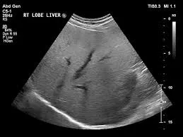
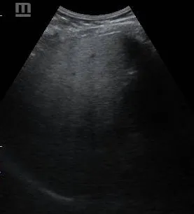
Normal findings
The normal liver demonstrates characteristic sonographic features that serve as the baseline for identifying pathology.
- Parenchymal Characteristics
- Homogeneous fine granular pattern
- Slightly hyperechoic compared to renal cortex
- Size
- Midclavicular line: ≤ 15.5cm
- Midline: ≤ 13 cm
- Margins
- Smooth, well-defined capsular surface
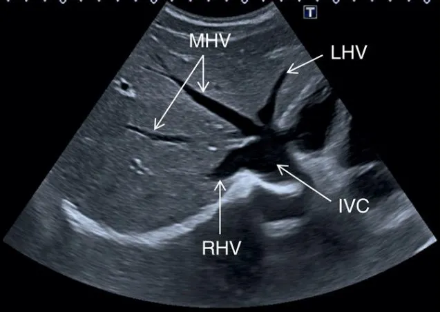
- Vasculature
- Should be patent with no evidence of thrombosis or dilatation
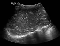
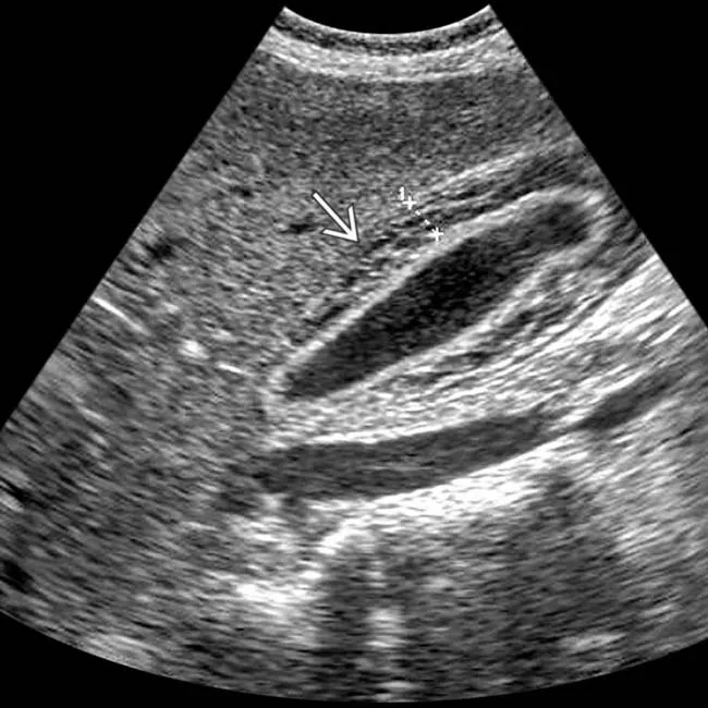
Common Pathological Findings
- Fatty liver
- Viral hepatitis
- Liver Cysts
- Peribiliary Cysts
- Autosomal Dominant Polycystic Disease
- Biliary Hamartomas (von Meyenburg Complexes)
- Liver abscess
- Hydatid Disease
Conclusion
Liver ultrasound remains an indispensable first-line imaging modality for evaluating hepatic pathology. A systematic approach assessing parenchymal echotexture, vascular patterns, and focal abnormalities enables accurate diagnosis of most hepatic conditions. While ultrasound excels as a screening tool, complementary imaging with contrast-enhanced ultrasound, CT, or MRI may be required for definitive characterization of certain lesions.
References
American College of Radiology. ACR Appropriateness Criteria® for Right Upper Quadrant Pain. 2021.
Dietrich CF, et al. EFSUMB Guidelines on Interventional Ultrasound. Ultraschall in Med. 2020.
Khalili K, et al. Sonographic features of hepatocellular carcinoma. J Clin Ultrasound. 2019.