Ultrasound Case: Hepatic Abscess
A 22-year-old male presents with high-grade fever and right upper quadrant (RUQ) abdominal pain.
Case Presentation
History
- Chief Complaint: High-grade fever (39.1°C) and right upper quadrant (RUQ) pain for 5 days.
- History of Present Illness:
- Progressive dull, aching RUQ pain.
- High-grade fevers accompanied by severe shaking chills.
- Marked anorexia and a 3kg weight loss over the past week.
- Denies any clinical history of jaundice or diarrhea.
- Past Medical History: Known history of asymptomatic gallstones, type 2 diabetes mellitus, and hypertension.
- Laboratory Findings:
- Leukocytosis: WBC 18.2 × 10³/µL with 90% neutrophils.
- Inflammatory Markers: CRP 156 mg/L.
- Liver Function Tests: ALT 85 U/L, AST 78 U/L, ALP 210 U/L (mild transaminitis and elevated alkaline phosphatase).
Physical Examination
- Abdomen: Significant tender hepatomegaly noted on palpation; localized guarding over the right hypochondrium.
- Signs: Positive physical Murphy’’s sign; no generalized rebound tenderness.
Clinical Suspicion: Given the combination of high-grade fever, leukocytosis, elevated CRP, and tender hepatomegaly in a diabetic patient, an acute intrahepatic infectious collection is highly suspected. Diagnostic ultrasound is indicated to differentiate acute cholecystitis from a hepatic abscess collection.
Ultrasound Findings
- Complex Hypoechoic Lesion: An ovoid, poorly defined intrahepatic mass is visualized within the right hepatic lobe, demonstrating irregular margins and low-level internal echo debris.
- Thick, Irregular Walls: The collection features an irregular, thick inflammatory pseudocapsule measuring approximately 4mm.
- Doppler Flow Characteristics: Color and power Doppler imaging show prominent peripheral hypervascularity around the wall (inflammatory rim), with a complete absence of internal vascularity within the fluid collection itself.
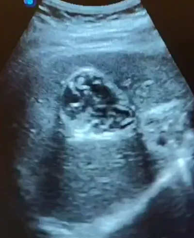
1. Right liver lobe: Large ovoid complex hypoechoic intrahepatic mass displaying highly irregular walls and non-homogeneous internal debris/echoes.
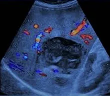
2. Color Doppler evaluation: Intense hypervascularity localized exclusively to the peripheral rim of the abscess wall with no detectable internal parenchymal flow.
Final Diagnosis: Pyogenic Hepatic Abscess
Confirmed by clinical markers, ultrasound-guided diagnostic fluid aspiration, and subsequent microbial culture growth revealing a Klebsiella pneumoniae infection.
Differential Diagnosis
- Necrotic Liver Metastasis: Can present as a large, complex hypoechoic hepatic lesion. Differentiated on ultrasound by the presence of irregular internal vascularity, a classic “target” or “bullseye” halo, and typically presenting in a patient with a known primary malignancy.
- Complex Hepatic Cyst: Features acoustic enhancement, but displays much thinner, smooth walls without a surrounding hypervascular inflammatory rim or systemic infective symptoms (high fevers, leukocytosis).
- Echinococcal (Hydatid) Cyst: Characterized by well-defined walls displaying a classic “wheel-spoke” pattern, internal daughter cysts, or a floating membrane (“water lily” sign); typically lacks acute pyogenic systemic signs.
- Hepatic Hematoma: Displays variable internal echotexture that evolves over time from anechoic to complex fluid-debris stages. It is avascular on Doppler imaging and characteristically associated with a clear history of localized abdominal trauma.