Ultrasound Case: Subcutaneous Lipoma of the Right Shoulder
Clinical History
A 31-year-old female presented with swelling over the posterior aspect of the right shoulder. A focused musculoskeletal ultrasound examination was performed to characterize the swelling, determine its location and dimensions, assess its vascularity, and evaluate the surrounding shoulder structures.
Ultrasound Findings
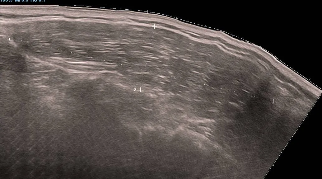
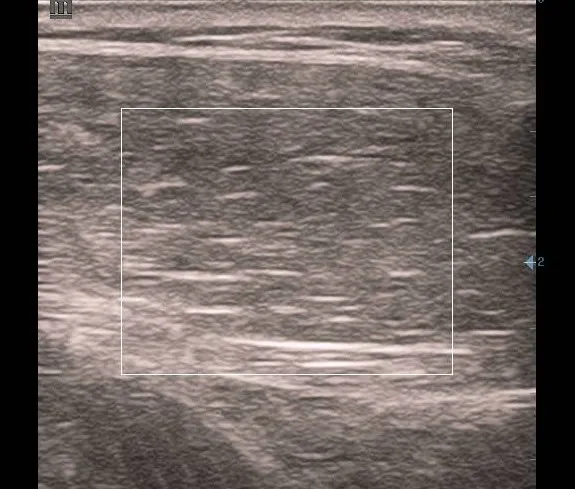
- A well-defined, oval-shaped, heterogeneous solid mass is identified within the subcutaneous tissues of the right shoulder region.
- The mass measures approximately 8.95 × 2.18 × 5.95 cm (longitudinal × anteroposterior × transverse dimensions, subject to the original scan orientation).
- No internal vascular flow is demonstrated on colour Doppler assessment.
- The right glenohumeral joint is well aligned.
- The humeral head and tuberosities demonstrate preserved cortical thickness.
- The right rotator cuff tendons demonstrate normal alignment and preserved fibrillar echotexture, with no focal masses or tendon tears.
- The rotator cuff tendon sheaths are free of fluid collections.
- No sonographic evidence of rotator cuff impingement is identified.
- The articular margins of the right acromioclavicular joint are regular, with maintained alignment.
- The surrounding shoulder muscles and fascial planes are reported as normal.
Diagnosis
Imaging impression: Well-defined subcutaneous soft-tissue mass of the right shoulder, suggestive of a lipoma.
The superficial location, oval shape, well-defined margins, and absence of detectable internal vascularity are compatible with a lipoma.
Histopathological confirmation may be obtained if the mass is excised or biopsied, particularly if clinical assessment or subsequent imaging raises concern.
Discussion
A lipoma is a benign tumour composed of mature adipose tissue. It is among the commonest benign soft-tissue masses and typically develops in the subcutaneous tissues of the trunk, neck, and extremities.
Lipomas often present as slowly enlarging, painless, palpable swellings. Depending on their size and location, they may cause discomfort, pressure symptoms, or cosmetic concerns.
Ultrasound Features of Lipoma
Typical sonographic features may include:
- A well-defined, oval or elongated superficial soft-tissue mass.
- Echogenicity that varies from hyperechoic to isoechoic or hypoechoic relative to the surrounding fat.
- Internal linear echogenic striations parallel to the long axis of the lesion in many cases.
- Little or no internal vascularity on colour Doppler examination.
- A location within the subcutaneous fat, although lipomas can also arise in deeper tissues.
The ultrasound appearance of lipomas can vary considerably. Some demonstrate homogeneous echotexture, whereas others are heterogeneous. Therefore, the diagnosis should be based on the overall imaging appearance and clinical findings rather than a single feature.
Interpretation in This Case
In this 31-year-old female, the mass is located in the subcutaneous tissue of the posterior right shoulder and measures approximately 8.95 cm in its longest reported dimension. Its well-defined oval configuration and lack of detectable internal Doppler flow support a benign superficial soft-tissue lesion, with lipoma being a leading consideration.
Nevertheless, the relatively large size and heterogeneous appearance warrant correlation with the clinical history, duration of swelling, growth pattern, consistency, and any associated pain. If the lesion is enlarging, painful, fixed, deep, or otherwise atypical, further assessment should be considered rather than assuming that it is a simple lipoma.
Differential Diagnosis
Important differential considerations include:
- Epidermoid cyst: A superficial, well-circumscribed lesion that may demonstrate internal debris and posterior acoustic enhancement. A punctum or connection to the skin may be present.
- Other benign soft-tissue masses: Including fibrous lesions and other benign mesenchymal tumours, whose appearances may overlap on ultrasound.
- Organized haematoma: May present as a heterogeneous mass, particularly following trauma or in patients with bleeding risk factors. Clinical history and interval changes are helpful.
- Vascular malformation: May present as a soft-tissue swelling, although Doppler assessment often demonstrates vascular channels or flow. Very slow-flow lesions may require careful evaluation.
- Atypical lipomatous tumour or other soft-tissue neoplasm: Less common considerations, particularly when a lesion is enlarging, deep, painful, or has suspicious imaging characteristics.
Management and Recommendations
Management depends on the clinical presentation, lesion size, growth pattern, and degree of diagnostic certainty.
- Clinical correlation: Establish the duration of the swelling, whether it is increasing in size, and whether there is associated pain, trauma, or functional limitation.
- Observation: May be appropriate for a clinically and radiologically typical lipoma that is stable and asymptomatic, following appropriate clinical assessment.
- Further imaging: Consider MRI for a large, deep, enlarging, painful, or indeterminate soft-tissue mass to better define its extent and internal characteristics.
- Specialist referral: A growing or suspicious soft-tissue mass should be referred for appropriate specialist assessment before any biopsy or excision.
- Histopathology: If the lesion is excised or biopsied, tissue examination can establish the definitive diagnosis.
Given the reported maximum dimension of approximately 8.95 cm and the heterogeneous appearance, clinical review is advisable to determine whether further imaging or specialist assessment is warranted.
Key Teaching Points
- Ultrasound is a useful first-line investigation for superficial soft-tissue swellings because it characterizes lesion location, margins, echotexture, and vascularity.
- A well-defined, superficial, oval mass with no detectable internal Doppler flow may suggest a lipoma, but these features are not diagnostic in isolation.
- Lipomas can have variable echogenicity and may appear heterogeneous on ultrasound.
- The size, depth, growth pattern, symptoms, and overall imaging characteristics should guide further assessment.
- Large or atypical soft-tissue masses should not automatically be labelled benign on the basis of ultrasound alone.
Clinical takeaway: The findings in this case are suggestive of a subcutaneous lipoma of the right shoulder. Clinical correlation and further evaluation, when indicated, are important to ensure appropriate management.