Ultrasound Case: Pelvic Inflammatory Disease (PID)
A 28-year-old female presents with lower abdominal pain and fever.
Case Presentation
History
- Chief Complaint: Lower abdominal pain x 5 days, fever (38.5°C), and vaginal discharge.
- History of Present Illness:
- Bilateral lower abdominal pain, worse on the right side.
- Increased yellowish vaginal discharge.
- Dyspareunia (pain during intercourse).
- No urinary symptoms.
- Past Medical History: Previous chlamydia infection (treated 2 years ago).
- Sexual History: Multiple partners, inconsistent condom use.
Physical Examination
- Abdomen: Tenderness in lower quadrants, rebound tenderness on the right.
- Pelvic Exam: Cervical motion tenderness (CMT), purulent cervical discharge.
- Vital Signs: Temp 38.5°C, HR 98, BP 110/70.
Clinical Suspicion: Given the history of STIs, multiple partners, fever, and cervical motion tenderness, PID is highly suspected. Ultrasound is requested to assess for complications such as tubo-ovarian abscess or pyosalpinx.
Ultrasound Findings
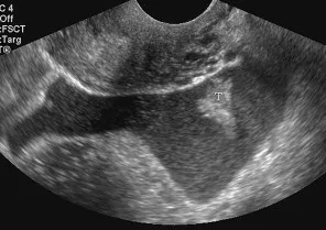
- Thickened, fluid-filled tubes: Exhibiting a “cogwheel” or “beads-on-a-string” appearance.
- Incomplete septations: Visualized within dilated tubes, strongly suggesting pyosalpinx.
- Free pelvic fluid: Present in the cul-de-sac.
- Tubo-ovarian complex: Ovary and fallopian tube are adherent but still distinguishable from one another.
- Hypervascularity: Marked increase in blood flow demonstrated on Doppler imaging due to active inflammation.
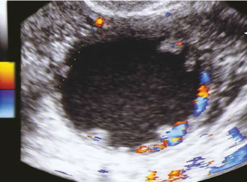
1. Right fallopian tube: Thickened, fluid-filled fallopian tube demonstrating incomplete septations and marked hypervascularity on Doppler imaging.
2. Free fluid: Anechoic to low-level echo complex free pelvic fluid in the cul-de-sac, indicative of purulent debris/pus.
Final Diagnosis: Pelvic Inflammatory Disease (PID) with Pyosalpinx
Confirmed based on matching clinical findings (fever, CMT, vaginal discharge) alongside key ultrasound features (dilated tubes, hypervascularity, and complex free fluid).
Differential Diagnosis
- Ectopic Pregnancy: Differentiate by positive β-hCG test and absence of fever; ultrasound typically reveals an adnexal mass displaying a hypervascular “ring of fire” on Doppler.
- Appendicitis: Presents with acute right lower quadrant (RLQ) pain and fever, but characteristically lacks cervical motion tenderness or purulent vaginal discharge.
- Ovarian Torsion: Marked by sudden, severe, unilateral pelvic pain; Doppler ultrasound demonstrates absent or severely compromised venous and arterial flow.
- Endometriosis: Presents with chronic pelvic pain and dysmenorrhea; ultrasound may display static ground-glass endometriomas, but patients lack acute systemic signs like fever.
- Diverticulitis: Features lower left quadrant (LLQ) pain and fever; distinct because gastrointestinal symptoms predominate.