Ultrasound Case: Pancreatic Pseudocyst
A 45-year-old male presents with history of recurrent pancreatitis, epigastric pain, and early satiety.
Case Presentation
History
- Chief Complaint: Dull epigastric pain and early satiety for 2 weeks.
- History of Present Illness:
- Persistent, dull epigastric pain characteristically radiating straight to the back.
- Notable early satiety accompanied by a documented 5kg weight loss over the last 2 months.
- No history of subjective fever, nausea, or vomiting during this episode.
- Relevant social history of alcohol-related acute pancreatitis (3 separate hospital admissions over the past year).
- Past Medical History: Documented chronic pancreatitis, type 2 diabetes mellitus, and severe hypertriglyceridemia.
- Laboratory Findings:
- Serum Amylase: 120 U/L (mildly elevated; normal range: 30–110 U/L).
- Serum Lipase: 85 U/L (mildly elevated; normal range: 7–60 U/L).
- Inflammatory Markers: WBC 9.8 × 10³/µL (normal), CRP 32 mg/L (mildly elevated).
Physical Examination
- Abdomen: Deep epigastric tenderness noted on palpation without guarding or signs of rebound; a firm, poorly defined, non-mobile mass is palpable within the epigastric region.
- General: No evidence of scleral icterus, peripheral jaundice, or ascites.
Clinical Suspicion: Given the patient’’s clear history of chronic pancreatitis alongside a palpable epigastric mass and early satiety, a mature post-pancontent fluid collection or a pancreatic pseudocyst is highly suspected. Diagnostic transabdominal ultrasound is indicated for structural confirmation.
Ultrasound Findings
- Anechoic Cystic Lesion: A large, well-defined, unilocular anechoic fluid collection is identified in the lesser sac, localized immediately anterior to the pancreatic body.
- Dimensional Profile: Measures approximately 8.5 x 6.2 cm in total size.
- Wall Integrity: Features a thin (1–2mm), smoothly demarcated fibrous pseudocapsule wall displaying a few thin, non-shadowing internal septations.
- Doppler Interrogations: Color and power Doppler demonstrate complete absence of internal vascularity or mural nodule flow.
- Background Pancreas: The surrounding pancreatic parenchyma shows classic signs of advanced chronic pancreatitis, characterized by a highly heterogeneous echotexture and multiple parenchymal calcifications.
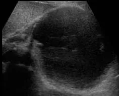
1. Epigastrium: Large, well-defined anechoic cystic fluid collection localized anterior to the pancreatic body with sharp acoustic enhancement.
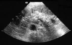
2. Pancreas bed: Highly heterogeneous, atrophic pancreatic parenchyma displaying bright echogenic parenchymal calcifications.
Final Diagnosis: Pancreatic Pseudocyst Complicating Chronic Pancreatitis
Confirmed by correlating the recurring clinical history of alcohol-related pancreatitis with sonographic evidence of a thin-walled, mature unilocular fluid collection localized to the lesser sac and subsequent abdominal CT cross-sectional correlation.
Differential Diagnosis
- Mucinous Cystic Neoplasm (MCN): Typically presents as a multilocular cystic lesion localized predominantly in the pancreatic body or tail of middle-aged females. Ultrasound demonstrates significantly thicker, irregular walls, thick internal septations, and occasional echogenic mural nodules with internal Doppler flow.
- Serous Cystadenoma (SCA): Characterized by a distinct microcystic or “honeycomb” appearance composed of numerous small cysts clustered together. Frequently displays a diagnostic central echogenic stellate scar or calcification block.
- Acute Pancreatic Abscess / Walled-Off Necrosis (WON): Presents with thick, highly irregular margins and prominent dependent fluid-debris levels or dirty internal gas echoes. Clinically distinguished by profound systemic septic parameters (high spiking fevers, toxic leukocytosis).
- Lymphoepithelial Cyst: A rare benign true cystic lesion of the pancreas that is typically found incidentally and is mostly asymptomatic. On ultrasound, it may mimic a pseudocyst but frequently contains complex internal echoes caused by gelatinous keratin debris.
Reference Articles
- Pancreas ultrasound technique and interpretation are covered in the current case content.