Ultrasound Case: Molar Pregnancy in a 16-Year-Old Female
Clinical History
A 16-year-old female patient presented with severe lower abdominal pain, abdominal fullness, and per-vaginal (PV) bleeding. A urine human chorionic gonadotropin (hCG) test was positive, indicating pregnancy.
Given the presenting symptoms and positive pregnancy test, pelvic ultrasound was indicated to evaluate the uterus and its contents, establish the likely cause of bleeding, and assess for possible complications.
Ultrasound Findings
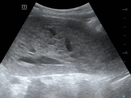
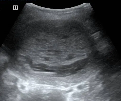
- An enlarged uterus containing heterogeneous intrauterine echogenic tissue. The uterus measured approximately 17.2 cm × 7.5 cm × 9.8 cm (longitudinal × anteroposterior × transverse).
- Multiple small cystic spaces within the intrauterine tissue, producing a characteristic vesicular or ‘snowstorm’ appearance.
- Absence of a normally developing embryo or fetus and no normal gestational sac indicating a complete hydatidiform mole.
- Variable vascularity within the abnormal intrauterine tissue on colour Doppler assessment.
- The ovaries demonstrated theca-lutein cysts, which occured likely due to elevated hCG levels.
- No free fluid was identified in the pelvis.
- The intrauterine content measured approximately 13.6 cm × 5.3 cm × 8.2 cm (volume = 316.2ml).
Diagnosis
The clinical presentation and imaging findings are highly suggestive of a hydatidiform mole (Complete molar pregnancy), presenting clinically with vaginal bleeding, severe lower abdominal pain, abdominal fullness, and a positive urine hCG test. Histopathological confirmation via fine needle aspiration cytology (FNAC) or biopsy was recommended.
Quantitative serum beta-hCG measurement and histopathological examination of evacuated tissue was recommended to support the diagnosis and for definitive classification.
Discussion
A hydatidiform mole is a form of gestational trophoblastic disease resulting from abnormal fertilisation and proliferation of placental trophoblastic tissue. It is classified into complete and partial hydatidiform moles.
Complete hydatidiform mole: Typically demonstrates diffuse abnormal trophoblastic proliferation, multiple cystic spaces within the uterine contents, and no embryo or fetus. Serum beta-hCG levels may be markedly elevated.
Partial hydatidiform mole: May demonstrate an abnormal gestational sac, a non-viable embryo or fetus, and focal cystic changes within an abnormal placenta. Its ultrasound appearance can overlap with that of a failed pregnancy.
Common clinical presentations include vaginal bleeding in early pregnancy, uterine enlargement greater than expected for gestational age, excessive nausea and vomiting, and occasionally symptoms associated with markedly elevated hCG levels.
Ultrasound is an important first-line investigation. However, early molar pregnancies may not show the classic vesicular appearance, and other conditions, including pregnancy loss with hydropic changes, can produce overlapping findings.
In this 16-year-old patient, the combination of vaginal bleeding, abdominal pain, abdominal fullness, and a positive urine hCG test raises concern for an abnormal pregnancy. The final diagnosis depends on correlation with ultrasound, quantitative serum beta-hCG, and histopathology.
Differential Diagnosis
Important differential considerations include:
- Incomplete or missed miscarriage: May present with vaginal bleeding and heterogeneous intrauterine material, but typically lacks the diffuse vesicular pattern of a complete mole.
- Partial hydatidiform mole: May resemble a failed pregnancy, with abnormal placental tissue and sometimes fetal structures.
- Retained products of conception: Heterogeneous intrauterine material may be present, particularly following a previous pregnancy loss or incomplete evacuation.
- Ectopic pregnancy: Must be considered in a patient with a positive pregnancy test and severe lower abdominal pain, particularly if no definite intrauterine pregnancy is identified.
- Other causes of early-pregnancy bleeding: Include subchorionic haemorrhage and other forms of early pregnancy loss.
Management
Suspected molar pregnancy requires prompt assessment by a gynaecology team, particularly in a patient presenting with severe pain and active vaginal bleeding.
Recommended management considerations include:
- Urgent clinical assessment: Evaluate haemodynamic stability, quantify bleeding, assess pain, and investigate for anaemia or other complications.
- Laboratory investigations: Obtain quantitative serum beta-hCG, full blood count, blood group and Rh status, and other investigations guided by the clinical findings.
- Confirmatory evaluation: Review the pelvic ultrasound findings and consider repeat imaging or specialist assessment if the diagnosis remains uncertain.
- Definitive treatment: When a hydatidiform mole is confirmed, uterine evacuation is generally performed under specialist care, with appropriate preparation and monitoring.
- Histopathology: Submit evacuated tissue for histological examination to confirm the diagnosis and distinguish complete from partial mole.
- Follow-up: Arrange serial quantitative beta-hCG monitoring until the recommended criteria for resolution are met. Persistent or rising hCG requires specialist evaluation for persistent gestational trophoblastic disease.
Because the patient is 16 years old, care should also include age-appropriate counselling, safeguarding assessment where indicated, and sensitive support for the patient.
Key Teaching Points
This case highlights the importance of considering molar pregnancy in a patient presenting with vaginal bleeding, abdominal pain, abdominal fullness, and a positive pregnancy test.
The key ultrasound teaching point is the recognition of abnormal intrauterine tissue containing multiple small cystic spaces, which may suggest a hydatidiform mole. However, not all molar pregnancies demonstrate the classic appearance, particularly in early gestation.
Clinical correlation is essential: A positive urine hCG test alone does not establish a molar pregnancy. Ultrasound findings, quantitative serum beta-hCG, clinical assessment, and histopathological examination help establish the diagnosis and guide appropriate management.
Severe lower abdominal pain and vaginal bleeding in early pregnancy require urgent assessment to exclude haemorrhage, ectopic pregnancy, and other potentially life-threatening complications.