Ultrasound Case: Ectopic Pregnancy
A 28-year-old female presents with right lower quadrant pain and vaginal spotting.
Case Presentation
History
- Chief Complaint: Right lower quadrant (RLQ) abdominal pain and vaginal spotting.
- History of Present Illness:
- 6 weeks since last normal menstrual period (LMP).
- Positive home qualitative urine pregnancy test.
- Sharp, non-radiating RLQ pain for 2 days, progressively worsening.
- Light, intermittent vaginal bleeding x 3 days.
- Denies fever, chills, or urinary track symptoms.
- Past Medical History: Previous clinical history of Pelvic Inflammatory Disease (PID) treated 2 years ago. Nulliparous (no prior pregnancies).
- Initial Labs: Quantitative serum β-hCG is 2,450 mIU/mL; progesterone is 8 ng/mL.
Physical Examination
- Vital Signs: Temp 36.8°C, HR 88, BP 120/80.
- Abdomen: Focused tenderness noted over the right lower quadrant with mild localized guarding; no clinical signs of generalized rebound tenderness.
- Pelvic Exam: Exquisite right adnexal tenderness and mild cervical motion tenderness (CMT); small amount of dark red blood visualized in the vaginal vault.
Clinical Suspicion: Given the documented history of pelvic inflammatory disease, an acute drop in progesterone with a serum β-hCG level well above the discriminatory zone (2,450 mIU/mL), and unilateral pelvic pain, an ectopic pregnancy is highly suspected. Emergency pelvic ultrasound is mandatory to localize the gestation sac.
Ultrasound Findings
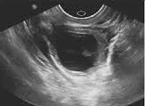
- Extrauterine Gestation Sac: Visualized in the right adnexa, distinct from the ovary, containing a well-defined early fetal pole without definitive cardiac activity.
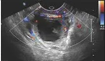
- Ring of Fire Sign: Color Doppler imaging demonstrates prominent, low-resistance peripheral hypervascular flow encircling the extrauterine gestation sac.
- Empty Uterus: Complete absence of a normal intrauterine pregnancy (IUP) or true gestational sac within the endometrial cavity; thin, reactive decidual reaction noted.
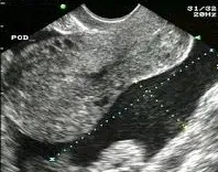
- Hemoperitoneum: Moderate collection of complex free fluid with low-level internal echoes visualized within the rectouterine space (pouch of Douglas).
Final Diagnosis: Right Adnexal Ectopic Pregnancy (Tubal)
Confirmed based on matching clinical markers (amenorrhea, vaginal bleeding, high serum β-hCG) alongside key diagnostic ultrasound features including an empty uterine cavity, an extrauterine adnexal gestation sac displaying a classic ‘ring of fire’ sign, and free pelvic hemoperitoneum.
Differential Diagnosis
- Corpus Luteum Cyst: Frequently adjacent to or inside the ovary, demonstrating a thick hypervascular wall on Doppler. However, it displays a classic eccentric cystic look, lacks an internal embryonic pole/yolk sac, and is associated with normal tracking intrauterine pregnancies.
- Hemorrhagic Ovarian Cyst: Ultrasound reveals a complex adnexal mass with characteristic fine reticular internal echoes (fishnet appearance) and a retractile clot. Crucially, it shows no peripheral Doppler flow in the clot itself and is completely avascular.
- Ovarian Torsion: Characterized by sudden, severe, unilateral pelvic pain. Sonography demonstrates a markedly enlarged, edematous ovary with peripheralized follicles; color and spectral Doppler reveal absent or significantly compromised venous and arterial flow profiles.
- Pelvic Inflammatory Disease (PID): Presents with bilateral lower abdominal pain, purulent discharge, and fever. Ultrasound features thickened, fluid-filled tubes (pyosalpinx) and adherent adnexal complexes, but is definitively ruled out by a positive serum β-hCG.
- Appendicitis: Presents with acute right lower quadrant pain and focal tenderness at McBurney’’s point. Ultrasound demonstrates a non-compressible, blind-ending tubular structure measuring >6mm in outer diameter in the RLQ; presents with a non-gravid uterus.