Renal Ultrasound
Renal ultrasound is a fundamental imaging modality for evaluating kidney pathology due to excellent visualization of the renal parenchyma and collecting system, real-time assessment of blood flow with Doppler, the absence of ionizing radiation or nephrotoxic contrast, and bedside capability for critically ill patients. However, it is operator-dependent, has limited evaluation of overall renal function, and can be restricted in obese patients or those with abundant bowel gas.
Renal Anatomy
Normal Kidney Structure
- Cortex: Hypoechoic to the liver or spleen and contains glomeruli.
- Medullary pyramids: More hypoechoic than the cortex.
- Sinus fat: A hyperechoic central area containing vessels and the collecting system.
- Pelvicalyceal system: Normally not distended.
Normal Measurements
- Length: 9-12 cm in adults.
- Cortical thickness: 7-10 mm.
- Parenchymal thickness: 15-20 mm.
- Resistive Index (RI): <0.7 in the main renal artery.
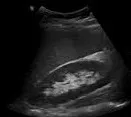
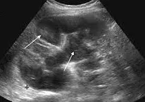
Demonstration of normal renal anatomy: cortex, medullary pyramids, and sinus fat.
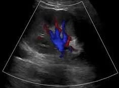
Color Doppler showing normal renal artery and vein flow at the hilum.
Clinical Indications
- Common Indications: Evaluation of flank or abdominal pain, hematuria, renal function impairment, hydronephrosis, and suspected renal stones[cite: 1].
- Clinical Scenarios: Includes management of renal colic, pyelonephritis, renal failure, and hypertension (evaluating for renal artery stenosis)[cite: 1].
Scanning Technique
- Preparation: While fasting is not strictly required, it helps reduce bowel gas, and a full bladder assists in evaluating the distal ureters.
- Equipment: Use a curvilinear transducer (2-5 MHz) for adults and adjust depth to include the entire kidney and surrounding structures.
- Approach: Perform a systematic survey using longitudinal and transverse views, utilizing the liver or spleen as acoustic windows, and use Doppler for vascular assessment.
Pathological Findings
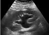
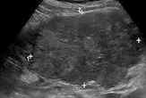
Grade 2 hydronephrosis showing dilated pelvis and calyces.
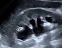
Renal calculus showing an echogenic focus with posterior acoustic shadowing.
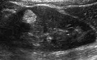
Acute pyelonephritis showing focal hypoechoic area with loss of corticomedullary differentiation.
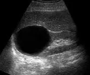
Simple renal cyst presenting as an anechoic lesion with posterior acoustic enhancement.
Well-circumscribed, hyperechoic fat-containing mass.
Heterogeneous solid mass distorting the renal contour.
References
- American College of Radiology (ACR). (2023). ACR Appropriateness Criteria® Renal Failure. Journal of the American College of Radiology, 20(1S), S78-S92.
- Rumack, C. M., et al. (2023). Diagnostic Ultrasound (6th ed.). Elsevier.
- European Federation of Societies for Ultrasound in Medicine and Biology (EFSUMB). (2022). Guidelines on Renal Ultrasound. Ultraschall in der Medizin, 43(3), 261-279.
- O’Neill, W. C. (2023). Renal Ultrasound. In: Johnson, R. J., et al. (Eds.), Comprehensive Clinical Nephrology (7th ed., pp. 112-128). Elsevier.
- African Society of Uroradiology (ASUR). (2023). Consensus Guidelines on Renal Ultrasound in Tropical Settings. African Journal of Radiology, 28(1), 45-60.