Periportal Cuffing on Ultrasound: Meaning, Causes and Clinical Significance
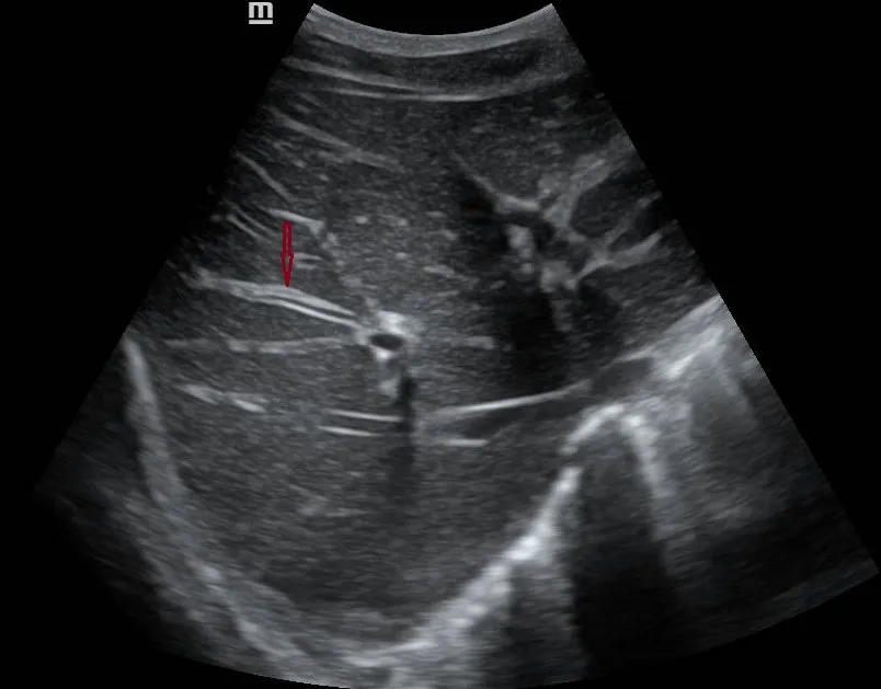
Periportal cuffing describes increased visibility or altered echogenicity surrounding the portal venous branches on liver ultrasound. Appearing as echogenic tissue or a halo-like rim around portal structures, it reflects changes in the connective tissue or fluid dynamics within the portal tracts.
While it is a prominent visual feature, periportal cuffing is a descriptive imaging finding—not a standalone diagnosis. It must always be interpreted in conjunction with patient history, laboratory findings, and overall hepatobiliary appearance.
Ultrasound Appearance
When evaluating the liver, systematically assess the portal tracts across multiple segments and describe the visual parameters without prematurely assigning an etiology:
- Echogenicity Pattern: Increased echogenicity or a cuff-like rim encircling portal branches (classically part of the “starry sky” appearance in severe diffusely hypoechoic liver parenchyma).
- Distribution: Determine whether the cuffing is focal or diffuse, symmetric or irregular.
- Associated Liver Findings: Note parenchymal echotexture changes, hepatomegaly, ascites, or signs of biliary dilatation.
- Biliary Tree: Evaluate whether intrahepatic and extrahepatic bile ducts appear normal, dilated, or demonstrate wall thickening.
If the finding is subtle or uncertain, document imaging limitations and request review by a senior radiologist.
Possible Causes and Differential Considerations
Periportal echogenicity is nonspecific. Differential diagnoses depend heavily on the clinical context and overall ultrasound pattern:
-
Acute Systemic Illness or Suspected Hepatitis
- Considerations: Inflammatory changes or acute periportal edema are common contributors, though not diagnostic on their own.
- Next Steps: Correlate with clinical symptoms, viral markers, and liver enzyme panels (ALT/AST).
-
Known Inflammatory Bowel Disease (IBD)
- Considerations: Reported in literature as an associated finding; however, it does not independently establish IBD-related liver disease.
- Next Steps: Review medication history, liver function tests, and hepatobiliary symptoms. Coordinate clinical assessment with gastroenterology.
-
Cholestatic Pattern or Abnormal Bile Ducts
- Considerations: Evaluate for underlying biliary disease, including early stricturing or biliary obstruction.
- Next Steps: Measure ductal caliber and morphology. Magnetic Resonance Cholangiopancreatography (MRCP) or further testing may be warranted.
-
Systemic Fluid overload or Inflammatory States
- Considerations: Periportal edema due to aggressive fluid resuscitation, congestive heart failure, or blunt abdominal trauma.
- Next Steps: Interpret alongside systemic hemodynamics, fluid balance, and trauma workup.
-
Regional/Infectious Etiologies
- Considerations: In endemic regions across Africa, consider parasitic infections such as Schistosomiasis (Symmers’ pipe-stem fibrosis), typhoid fever, or severe malaria.
- Next Steps: Correlate with travel history, blood smears, and serological testing.
This differential is a practical framework rather than an exhaustive list. Periportal cuffing alone should never be used to make a definitive diagnosis.
Reported Association with Inflammatory Bowel Disease
A notable case series published in Inflammatory Bowel Diseases highlighted periportal cuffing in patients with IBD, proposing that altered immune-cell trafficking along the gut–liver axis might drive periportal changes. In these reported cases, MRCP did not demonstrate classic features of Primary Sclerosing Cholangitis (PSC), and contrast ultrasound ruled out periportal abscesses.
However, this report is hypothesis-generating rather than diagnostic. A small series cannot prove causation or establish periportal cuffing as a reliable biomarker for IBD flare-ups.
Suggested Reporting Approach
Radiologists should clearly describe the finding while avoiding over-interpretation.
Example Template Wording:
“Mild periportal echogenicity/cuffing is demonstrated throughout the liver parenchyma. The finding is non-specific and may reflect periportal edema or inflammatory changes. Clinical correlation with liver function tests and patient history is recommended.”
FAQ & Clinical Pearls
Is periportal cuffing the same as periportal edema?
Not strictly. “Cuffing” describes the visual ultrasound pattern, whereas edema is one of several underlying histological causes.
Does periportal cuffing confirm Primary Sclerosing Cholangitis (PSC)?
No. Periportal cuffing alone cannot diagnose PSC. Diagnosing PSC requires clinical evaluation and characteristic biliary tree beaded changes, typically confirmed via MRCP.
What details should be included in the formal report?
Specify the distribution (focal vs. diffuse), degree of echogenicity, presence/absence of biliary ductal dilation, gallbladder wall thickness, and recommend clinical/laboratory correlation.
References
- Inflammatory Bowel Diseases Journal (2010): Periportal Cuffing in Inflammatory Bowel Diseases: Mystery of Stars and Stripes. DOI: 10.1002/ibd.21165
- Rumack, C. M., & Levine, D. (2023): Diagnostic Ultrasound (6th ed.). Elsevier — Chapter on Hepatobiliary Sonography.
- African Society of Radiology (ASR) (2023): Abdominal Ultrasound Protocols in Endemic Hepatobiliary Conditions. African Journal of Medical Imaging, 14(1), 42–55.