Neonatal Femoral Fracture After Vaginal and Caesarean Delivery: The Role of Ultrasound in Diagnosis
Introduction
Neonatal femoral fracture is an uncommon but clinically important birth injury involving the thigh bone (femur) of a newborn[cite: 1]. Although rare, it can occur during both vaginal delivery and Caesarean section, particularly when the baby is in a breech presentation or delivery requires difficult extraction[cite: 1].
Early recognition is important because a femoral fracture may initially present with subtle findings, such as reduced movement of one leg, localized swelling, tenderness, or discomfort during handling. In some cases, swelling becomes apparent only several days after birth, delaying diagnosis.
Ultrasound is a valuable imaging tool for assessing suspected neonatal fractures. It provides real-time visualization of the bone surface, surrounding soft tissues, and early fracture healing without exposing the infant to ionizing radiation. However, conventional radiography remains an important diagnostic examination, particularly for assessing fracture alignment and the overall pattern of injury[cite: 1].
What Is a Neonatal Femoral Fracture?
A neonatal femoral fracture is a break in the femur occurring before, during, or shortly after birth. Birth-related fractures commonly involve the femoral shaft, although injuries around the proximal or distal femoral growth plate can also occur.
Femoral shaft fractures may be transverse, oblique, or spiral in configuration. Spiral and short-oblique fractures can result from rotational or torsional forces applied to the limb during difficult delivery.
Birth-related femoral fractures are very rare[cite: 1]. Published literature reports an estimated incidence of approximately 0.013%–0.089% of newborns, although reported rates vary between studies and populations[cite: 1].
Relationship Between Mode of Delivery and Femoral Fracture
1. Vaginal Delivery
Vaginal delivery is a recognized setting for neonatal femoral fracture, particularly during breech presentation.
In a breech delivery, the baby’s buttocks or feet emerge first. If the legs are difficult to deliver, excessive traction or rotation may transmit mechanical forces to the femur, potentially resulting in a fracture.
Potential risk factors include:
- Breech presentation, particularly when extraction is difficult.
- Prematurity or low birth weight.
- Difficult extraction of the baby’s legs or pelvis.
- Multiple pregnancy.
- Underlying bone fragility or skeletal disorders.
Femoral fractures can also occur during cephalic vaginal delivery, although this is uncommon.
2. Caesarean Section
Caesarean delivery generally reduces some forms of mechanical birth trauma associated with vaginal breech delivery, but it does not eliminate the risk of femoral fracture.
Fractures have been reported during Caesarean section, especially when delivering a breech baby or when extraction is difficult[cite: 1]. Restricted space for manoeuvring the infant, forceful traction, rotation, or levering of a limb during extraction may contribute to injury.
Therefore, the occurrence of a neonatal femoral fracture following Caesarean delivery does not automatically indicate improper technique[cite: 1]. The circumstances of delivery, clinical findings, imaging results, and other relevant risk factors should be evaluated together[cite: 1].
Important Clinical Point: Neither vaginal delivery nor Caesarean section completely eliminates the possibility of neonatal femoral fracture. The risk depends on the presentation of the fetus, the difficulty of extraction, and other maternal, fetal, and procedural factors.
Clinical Presentation
The clinical signs of a neonatal femoral fracture may be apparent immediately after delivery or develop over the following days.
Common findings include:
- Reduced spontaneous movement of the affected leg.
- Localized swelling or tenderness of the thigh.
- Pain or crying when the limb is handled.
- Abnormal positioning or apparent deformity of the leg.
- Crepitus at the fracture site, although this may be absent.
- Asymmetry between the two lower limbs.
In some newborns, the fracture may not be suspected until progressive swelling or reduced limb movement becomes noticeable.
A careful examination should assess both lower limbs, including their movement, perfusion, skin condition, and any associated injuries.
Ultrasound Assessment of Neonatal Femoral Fracture
Ultrasound is particularly useful in neonatal musculoskeletal imaging because the infant’s bones are small and parts of the skeleton, especially the epiphyses, remain cartilaginous.
High-frequency linear transducers provide detailed images of the superficial femoral cortex and surrounding soft tissues. Ultrasound can be performed at the bedside, requires no ionizing radiation, and generally does not require sedation.
Ultrasound Technique
A focused examination should include the following steps:
- Position the newborn comfortably, minimizing unnecessary movement of the affected limb.
- Use a high-frequency linear transducer and apply a suitable amount of ultrasound gel.
- Examine the femur in longitudinal and transverse planes.
- Scan the accessible anterior, medial, and lateral surfaces of the bone.
- Compare the affected femur with the contralateral side when appropriate.
- Assess the surrounding soft tissues for swelling, hematoma, or other abnormalities.
- Document the fracture location, cortical disruption, displacement, angulation, and any evidence of healing.
The examination should be performed gently. The suspected fracture should not be stressed or manipulated to demonstrate movement.
Characteristic Ultrasound Findings
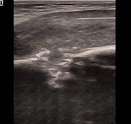
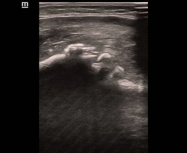
- Disruption of Cortical Continuity: The normal femoral cortex appears as a smooth, continuous echogenic line with posterior acoustic shadowing. At the fracture site, this line may be interrupted, irregular, or discontinuous.
- Cortical Step-Off: A break in the cortex may produce an abrupt step or offset between the proximal and distal bone fragments.
- Angulation or Displacement: The bone fragments may demonstrate abnormal alignment, with angulation, translation, or overlap depending on the fracture configuration.
- Periosteal Elevation: The periosteum may appear elevated or displaced at the fracture site. This finding can be particularly useful when assessing injuries in young infants.
- Adjacent Soft-Tissue Changes: Localized soft-tissue swelling or a small hematoma may be visible around the fracture.
- Callus Formation: During healing, new bone formation may appear as irregular echogenic tissue around the fracture site. Progressive callus formation and improvement in alignment may be observed on follow-up imaging.
These features must be interpreted together with the clinical history and examination. Ultrasound findings can be subtle, especially in fractures involving the growth plate or predominantly cartilaginous structures.
Ultrasound Versus X-Ray
Both ultrasound and radiography have important roles in evaluating neonatal femoral fractures.
| Feature | Ultrasound | X-Ray |
|---|---|---|
| Ionizing Radiation | None | Uses ionizing radiation |
| Bedside Examination | Possible | Portable examination possible |
| Cortical Disruption | Often well demonstrated | Usually well demonstrated |
| Cartilage and Periosteum | Particularly useful | May be difficult to assess |
| Fracture Alignment | Can demonstrate angulation and displacement | Provides an established overview of alignment |
| Soft-Tissue Assessment | Good | Limited |
| Limitations | Operator-dependent; some fracture patterns may be missed | Early or predominantly cartilaginous injuries may be difficult to identify |
Radiography is commonly used to confirm and characterize a suspected femoral shaft fracture. Ultrasound is a valuable complementary examination and may be particularly helpful when the radiographic findings are inconclusive or when a distal femoral physeal injury is suspected.
A normal ultrasound does not reliably exclude every neonatal fracture. If clinical suspicion remains high, additional imaging and specialist assessment are appropriate.
Differential Diagnoses
A swollen or poorly mobile leg in a newborn does not always indicate a femoral fracture. Relevant differential diagnoses include:
- Femoral Physeal Injury: Damage around the growth plate may be difficult to identify on conventional radiographs because the epiphysis is incompletely ossified.
- Septic Arthritis: May cause reduced limb movement, pain, and swelling around the hip or knee.
- Osteomyelitis: Bone infection may produce tenderness, swelling, and reduced movement.
- Congenital Femoral Abnormalities: Femoral shortening or bowing may be present from birth.
- Skeletal Fragility Disorders: Conditions such as osteogenesis imperfecta may predispose an infant to fractures.
- Soft-Tissue Injury: Bruising, hematoma, or muscle injury may cause swelling without a fracture.
Clinical history, examination, imaging, and appropriate laboratory investigations help distinguish these conditions.
Management and Follow-Up
Once a femoral fracture is suspected or confirmed, the newborn should receive prompt assessment by a pediatric orthopedic or appropriately experienced neonatal team.
Most uncomplicated birth-related femoral shaft fractures can be managed conservatively[cite: 1]. Treatment depends on the fracture location, displacement, angulation, associated injuries, and the infant’s overall condition[cite: 1].
Management may include:
- Gentle handling and appropriate limb immobilization.
- A Pavlik harness, suitable strapping, or casting when indicated by the treating specialist.
- Analgesia appropriate for the newborn.
- Clinical and imaging follow-up to assess healing and alignment.
- Investigation for underlying bone fragility when the fracture pattern, clinical history, or other findings warrant it.
Ultrasound may help document callus formation during healing, potentially reducing the need for some repeat radiographs in selected cases. Nevertheless, the follow-up imaging method should be determined by the treating team.
Neonatal bones have substantial remodelling potential, and most isolated birth-related femoral fractures heal well with appropriate care[cite: 1]. Long-term outcomes are generally favourable, although follow-up is important to assess limb alignment, length, and function[cite: 1].
Prevention and Clinical Considerations
Prevention focuses on careful obstetric assessment and appropriate delivery techniques.
Important considerations include:
- Identifying breech presentation and other factors that may complicate delivery.
- Planning delivery according to the clinical circumstances and established obstetric guidance.
- Using appropriate manoeuvres during delivery or extraction.
- Avoiding unnecessary force or excessive traction on the fetal limbs.
- Examining the newborn promptly when limb movement is reduced or swelling is present.
- Arranging early imaging when a birth-related fracture is suspected.
A neonatal femoral fracture can occur despite appropriate obstetric care. The presence of a fracture alone is insufficient to determine its mechanism or establish that an error occurred during delivery.
Conclusion
Neonatal femoral fracture is a rare but significant birth injury that can occur following both vaginal delivery and Caesarean section[cite: 1]. Breech presentation and difficult extraction are important clinical considerations, but neither mode of delivery completely eliminates the risk[cite: 1].
Ultrasound is a useful, radiation-free imaging technique for identifying cortical disruption, fracture displacement, periosteal elevation, surrounding soft-tissue changes, and callus formation. Its role is particularly valuable in neonatal musculoskeletal assessment, where incompletely ossified structures may complicate radiographic interpretation.
For suspected femoral fractures, ultrasound should be integrated with clinical assessment and radiography when indicated[cite: 1]. Early diagnosis, appropriate immobilization, and specialist follow-up generally result in favourable outcomes[cite: 1].
References
- McDonald TC, Higdon LC, Cutchen AW. Neonatal Birth Fractures. Review article. Available at: PMC Article[cite: 1]
- Musculoskeletal imaging in neonates: use of ultrasound. Review article. PubMed. Available at: PubMed Entry
- Birth-related femoral fracture in newborns: risk factors and management. Ethiopian Journal of Health Sciences / case-series literature. Available at: PMC Article
- Liu JL, Zhang L, Qiu RX. Ultrasound Instead of X-Ray to Diagnose Neonatal Fractures: A Feasibility Study Based on a Case Series. Frontiers in Pediatrics. 2022;10:847776. Available at: Frontiers Journal